
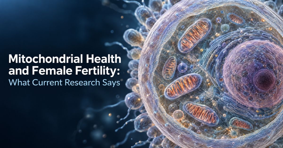
Every egg cell a woman’s body prepares for ovulation requires an extraordinary amount of energy. That energy comes from mitochondria — structures inside cells that convert nutrients into the chemical fuel that drives nearly every biological process. In reproductive biology, mitochondria are not a minor detail. They are central to whether an egg matures properly, fertilises successfully, and supports the earliest stages of embryonic development.
Over the past decade, mitochondrial function has moved from the margins of fertility research to one of its most active frontiers. Multiple peer-reviewed studies published between 2020 and 2026 have examined how mitochondrial health influences oocyte quality, what happens when mitochondrial function declines with age, and whether targeted interventions can support the cellular environment in which eggs develop.
This article explains the biology, reviews the evidence, and distinguishes between what is established, what is promising, and what remains unproven.
What Are Mitochondria and Why Do They Matter?
Mitochondria are membrane-bound organelles found in nearly every cell in the human body. Their primary function is to produce adenosine triphosphate (ATP) — the molecule that cells use as their main energy currency. This process, known as oxidative phosphorylation, takes place within the inner mitochondrial membrane and involves a series of enzyme complexes called the electron transport chain.
Beyond energy production, mitochondria participate in several other processes relevant to cellular health: calcium signalling, regulation of programmed cell death (apoptosis), and maintenance of the cell’s internal oxidation–reduction balance. When mitochondria function efficiently, cells have the energy and molecular signals they need to carry out their specialised tasks. When mitochondrial function is impaired, cellular processes slow down, errors accumulate, and the cell’s ability to perform its role is compromised.
What makes mitochondria particularly relevant to reproductive biology is the sheer scale of their presence in egg cells. A mature human oocyte contains approximately 100,000 to 600,000 mitochondria — far more than any other cell type in the body. This is not coincidental. The egg must store enough energy to power its own final stages of maturation, the process of fertilisation, and the first several days of embryonic cell division before the embryo’s own mitochondria begin to replicate.
Why Mitochondria Are Critical in Egg Development
Oocyte maturation is an energy-intensive process. During the final stages before ovulation, the egg must complete meiosis — a specialised form of cell division that reduces the chromosome number from 46 to 23, preparing it for fusion with a sperm cell. This process requires precise chromosome alignment, accurate spindle formation, and the extrusion of a polar body, all of which depend on adequate ATP supply.
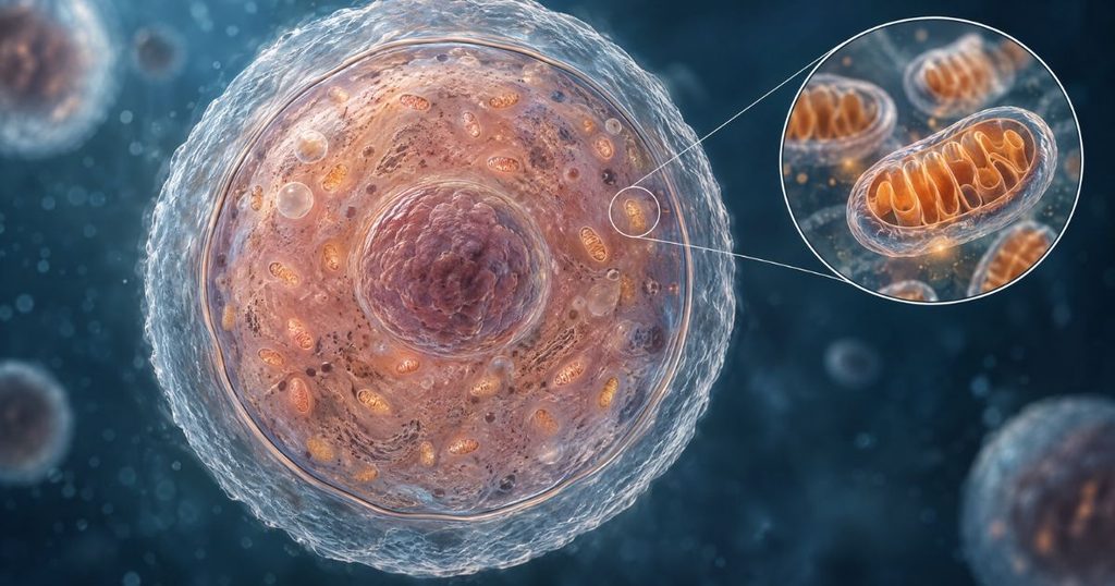
Research published in the Journal of Ovarian Research (2025) confirmed that when mitochondrial dysfunction occurs in the oocyte, reduced metabolic efficiency impairs both nuclear and cytoplasmic maturation. The consequences can include abnormal spindle assembly, chromosome misalignment, and ultimately aneuploidy — the wrong number of chromosomes in the resulting embryo. Aneuploidy is the most common cause of early miscarriage and failed implantation.
After fertilisation, the early embryo relies entirely on the mitochondria it inherited from the egg. Sperm contribute virtually no mitochondria to the embryo. This means the egg’s mitochondrial reserves must sustain the first several rounds of cell division — from zygote through morula to blastocyst — before the embryo’s own mitochondrial biogenesis begins. If those reserves are insufficient, embryo development stalls.
A 2024 editorial in Frontiers in Endocrinology noted that mitochondrial DNA copy number in oocytes and cumulus granulosa cells has been investigated as a potential biomarker of reproductive potential, reflecting the direct connection between mitochondrial health and fertility outcomes.
Mitochondrial Function, Egg Quality, and Reproductive Ageing
Egg quality, ovarian reserve, and mitochondrial function are related but distinct concepts. Ovarian reserve refers to the number of remaining follicles in the ovaries, typically estimated through AMH levels and antral follicle counts. Egg quality refers to the developmental competence of individual oocytes — their ability to mature, fertilise, and produce viable embryos. Mitochondrial function is one of several biological factors that influences egg quality, alongside chromosomal integrity, cytoplasmic organisation, and the quality of surrounding granulosa cells.
With advancing age, all three tend to decline, but they do not decline in lockstep. A woman may have a low ovarian reserve but still produce eggs of reasonable quality. Conversely, a woman with an adequate follicle count may produce eggs with compromised mitochondrial function. This is why fertility assessments that focus only on egg number can be misleading.
What happens to mitochondria as reproductive ageing progresses? Research reviewed in Frontiers in Endocrinology (2024) and Human Reproduction Update (2025) identifies several age-related changes:
Reduced mitochondrial membrane potential. The inner membrane potential is essential for ATP production. As it declines, less energy is available for oocyte maturation.
Increased reactive oxygen species (ROS) production. Ageing mitochondria become less efficient and produce more byproduct oxidative molecules, which can damage DNA, proteins, and lipids within the cell.
Alterations in mitochondrial DNA. Both point mutations and deletions in mitochondrial DNA accumulate with age. A study in Human Reproduction Open (2025) found that specific mitochondrial DNA haplogroups were associated with differences in ovarian response, suggesting that mitochondrial genetics play a role in individual fertility variation.
Impaired mitochondrial quality control. Processes such as mitophagy (the selective removal of damaged mitochondria) and mitochondrial fusion and fission become less efficient, allowing damaged mitochondria to persist rather than being replaced.
These changes are gradual and begin well before they become clinically apparent. The acceleration after age 35 that fertility specialists commonly reference reflects, in part, the cumulative effect of mitochondrial decline reaching a threshold where it measurably affects oocyte competence.
Oxidative Stress and Female Reproductive Health
Oxidative stress occurs when the production of reactive oxygen species exceeds the cell’s antioxidant defences. In small quantities, ROS serve as signalling molecules involved in normal cellular processes, including aspects of follicular development. In excess, they cause indiscriminate damage to cellular components.
In the context of reproductive biology, oxidative stress has been linked to impaired oocyte maturation, DNA fragmentation, abnormal spindle formation, and reduced embryo viability. A 2017 review in Reproductive Biomedicine Online noted that excessive ROS can interfere with oocyte maturation and may inhibit in-vitro maturation processes.
Several factors contribute to oxidative stress in reproductive cells: advancing age, environmental toxin exposure, smoking, poor nutrition, chronic inflammation, and — notably — mitochondrial dysfunction itself. As mitochondria become less efficient, they produce more ROS as a byproduct of impaired electron transport. This creates a feedback loop: mitochondrial dysfunction increases oxidative stress, and oxidative stress further damages mitochondria.
It is important to note that the presence of oxidative stress does not automatically translate into clinical infertility, and reducing oxidative stress through antioxidant supplementation does not automatically improve pregnancy outcomes. The relationship is more nuanced than supplement marketing typically suggests. Antioxidant mechanisms address one contributing factor among several, and their clinical significance depends on the individual’s specific biological context.
What Does Research Say About NMN and Female Reproductive Health?
Nicotinamide mononucleotide (NMN) is a precursor to nicotinamide adenine dinucleotide (NAD+), a coenzyme involved in hundreds of metabolic reactions, including mitochondrial energy production and DNA repair. NAD+ levels decline with age across multiple tissues, and this decline has been implicated in various aspects of cellular ageing.

The hypothesis behind NMN supplementation in reproductive health is straightforward: by replenishing NAD+ levels, it may be possible to support mitochondrial function in ageing oocytes. The research supporting this hypothesis exists at several levels of evidence, and the distinctions between them matter.
Animal Studies
The strongest body of evidence comes from animal research. Studies by Bertoldo et al. (2020), Yang et al. (2020), and Ma et al. (2024) demonstrated that NMN supplementation in aged mice restored NAD+ levels in oocytes, improved oocyte maturation rates, enhanced mitochondrial membrane potential, and improved fertilisation and embryo development outcomes. A 2025 systematic review published in the Journal of Assisted Reproduction and Genetics examined seven high-quality preclinical studies and concluded that NMN supplementation shows consistent improvements in oocyte function across diverse biological stressors.
These findings are significant but must be contextualised. Mouse reproductive physiology shares important parallels with human reproduction, but the timescales, dosing, metabolic pathways, and clinical endpoints differ. Results in animal models do not automatically predict the same outcomes in humans.
Human In-Vitro Evidence
The most notable human-relevant study was published in the American Journal of Obstetrics and Gynecology (2025) by Ramírez-Martín et al. The researchers applied NMN supplementation to immature human oocytes from patients of advanced maternal age using a germinal vesicle (GV) rescue approach. NMN treatment improved NAD+ levels, promoted mitochondrial redistribution, and enhanced oocyte maturation competence. The authors concluded that their findings support further investigation of NMN’s clinical applications in female fertility, while emphasising that additional human studies are needed.
This is the strongest published evidence involving human oocytes, but it was conducted in vitro (in the laboratory, not inside the body) and did not measure pregnancy or live birth outcomes.
Early Clinical Observations
A retrospective analysis presented at the European Society of Human Reproduction and Embryology (ESHRE) 2025 meeting examined NMN pretreatment in young women with diminished ovarian reserve. The NMN-treated group showed improved follicle growth, higher fertilisation rates, and a higher pregnancy rate compared to controls. However, this was a retrospective analysis with a small sample size, not a randomised controlled trial, and it was presented as a conference abstract rather than a full peer-reviewed publication.
What This Evidence Does and Does Not Show
The evidence collectively supports the biological plausibility of NMN as a mitochondrial support compound in reproductive contexts. It does not yet establish proven clinical efficacy for improving pregnancy rates or live birth outcomes in humans through oral supplementation. This is an important distinction. Biological plausibility and preclinical promise are the beginning of the evidence pathway, not its conclusion.
Research on Trans-Resveratrol and Trans-Pterostilbene
Trans-resveratrol, a natural polyphenol found in grape skins and Japanese knotweed, has been studied for its effects on oocyte quality across multiple models. A systematic review from Anglia Ruskin University, encompassing 24 studies and over 9,500 participants, found evidence suggesting resveratrol can improve the quantity and quality of oocytes. Resveratrol activates the SIRT1 pathway — a cellular signalling mechanism involved in DNA repair and stress response — and has demonstrated antioxidant and anti-inflammatory properties in reproductive tissues. Most evidence remains preclinical, and the researchers emphasised the need for further human clinical trials to establish safe and effective dosing.
Trans-pterostilbene, a dimethylated analogue of resveratrol with reportedly greater bioavailability, was investigated in a 2025 study published in Aging. In aged mice, short-term pterostilbene ingestion improved implantation rates, while long-term supplementation in middle-aged mice enhanced implantation and live birth rates and increased the number of ovulated oocytes. At the cellular level, pterostilbene improved mitochondrial membrane potential and ATP levels in oocytes. Notably, unlike resveratrol, pterostilbene did not inhibit decidualisation in endometrial stromal cells — a finding the authors highlighted as a potential advantage for fertility applications. These results are from mouse models and require human validation.
Research on EGCG
Epigallocatechin gallate (EGCG), the primary polyphenol in green tea, has been studied for its antioxidant properties in reproductive contexts. A 2024 study in Cell Proliferation demonstrated that EGCG improved the quality of maternally aged oocytes in mice by reducing oxidative stress and supporting mitochondrial function. Earlier research had shown EGCG’s protective effects on oocytes exposed to environmental toxins and post-ovulatory ageing. A 2020 review in Biomedicine and Pharmacotherapy noted EGCG’s potential in protecting germ cells from damage through regulation of reactive oxygen species. As with other compounds reviewed here, the evidence is primarily from animal and in-vitro studies.
Lifestyle, Nutrition, and Evidence-Based Fertility Care
Mitochondrial health is not determined solely by supplementation. Several lifestyle and nutritional factors have established associations with reproductive health, supported by general medical consensus:
Balanced nutrition. Diets rich in antioxidants — including colourful vegetables, fruits, nuts, seeds, and whole grains — provide the micronutrients that support cellular defence mechanisms. No single food has been shown to reverse reproductive ageing, but adequate nutrition supports the cellular environment in which eggs mature.
Tobacco avoidance. Smoking is directly toxic to ovarian tissue and accelerates both ovarian reserve depletion and oxidative damage to oocytes. Cessation is among the most evidence-supported interventions for protecting reproductive health.
Moderate physical activity. Regular exercise supports metabolic health, insulin sensitivity, and hormonal balance — all relevant to reproductive function. Excessive exercise, however, can impair ovulation.
Sleep quality. Adequate sleep supports hormonal regulation, including the cyclical release of reproductive hormones. Chronic sleep disruption has been associated with menstrual irregularity and impaired follicular development.
Stress management. While the direct causal link between psychological stress and fertility outcomes is complex and debated, chronic stress elevates cortisol and can disrupt the hypothalamic-pituitary-ovarian axis.
Limiting alcohol consumption. Research associates heavy alcohol use with reduced fertility, though the threshold for harmful effects remains debated.
These recommendations are general health guidelines that apply broadly and are not specific to mitochondrial health. They represent the foundation upon which any targeted intervention, including supplementation, is built.
Understanding Mitochondrial Health Supplements
The growing research interest in mitochondrial function and reproduction has led to an expanding market of supplements positioned for fertility support. When evaluating any such product, readers and their healthcare professionals should consider several factors:
Ingredient evidence level. Has each ingredient been studied in the context of reproductive health specifically, or is the claim based on general antioxidant activity? What type of evidence exists — animal studies, in-vitro research, or human clinical trials?
Formulation vs. ingredient research. Research on individual ingredients does not automatically validate a specific formulation. The combination of ingredients, their dosages, bioavailability, and potential interactions within a finished product are separate questions from whether each ingredient has been studied individually.
Claims vs. evidence. Supplements in India are classified as nutraceuticals and are not required to demonstrate clinical efficacy in the same way as pharmaceutical drugs. Marketing claims should be assessed against the actual published evidence.
Medical supervision. Any supplement used in the context of fertility treatment — particularly alongside IVF, IUI, or hormonal protocols — should be discussed with a qualified fertility specialist who can assess the individual’s specific clinical situation.
Questions worth discussing with a healthcare professional include: What is my specific fertility diagnosis? Would mitochondrial support be relevant to my situation? Are there any interactions with my current medications or treatment protocol? What is the realistic evidence basis for the supplements I am considering?
Understanding MITOV
MITOV, developed by Surishi Pharmaceuticals, is positioned as India’s first mitochondrial optimiser formulated specifically for female fertility and subfertility support. It is a non-hormonal nutraceutical intended for use under medical supervision.
Its verified composition includes β-nicotinamide mononucleotide (NMN) at 425 mg — its primary active ingredient — alongside trans-resveratrol (25 mg), trans-pterostilbene (25 mg), simarouba (10 mg), Rabdosia rubescens (5 mg), and epigallocatechin gallate (EGCG, 2.5 mg).
As discussed in earlier sections, several of these ingredients have been studied individually in reproductive biology contexts. NMN has the most developed evidence base, primarily from animal studies and early human in-vitro research. Trans-resveratrol and trans-pterostilbene have demonstrated oocyte-protective effects in animal models, and EGCG has shown antioxidant activity relevant to oocyte quality in preclinical research.
It is important to note that clinical trials demonstrating that the complete MITOV formulation improves pregnancy rates, live birth outcomes, or IVF success rates in humans have not been published at the time of writing. The product’s positioning is based on the biological rationale of supporting mitochondrial function using ingredients with individual research backing, rather than on completed clinical trials of the finished product. Readers considering MITOV should discuss its use with their fertility specialist in the context of their individual treatment plan. Further information about MITOV’s composition and intended use is available on the MITOV product page (surishi.in/product/mitov/).
FAQs :
Q1. What role do mitochondria play in egg development?
Mitochondria produce the ATP (cellular energy) that eggs require for their final stages of maturation, fertilisation, and early embryonic cell division. A mature human egg contains far more mitochondria than any other cell type in the body — estimated between 100,000 and 600,000 — because it must store enough energy to sustain the embryo until its own mitochondrial replication begins.
Q2. Is mitochondrial function the same as egg quality?
No. Egg quality is a broader concept that encompasses chromosomal integrity, cytoplasmic organisation, and the developmental competence of the oocyte. Mitochondrial function is one important contributing factor to egg quality but does not define it entirely. A woman can have mitochondrial concerns alongside normal chromosomal health, or vice versa.
Q3. What is known about mitochondrial changes with reproductive ageing?
Research indicates that mitochondria in oocytes undergo several age-related changes: reduced membrane potential (less energy production), increased production of reactive oxygen species, accumulation of mitochondrial DNA mutations, and impaired quality-control mechanisms. These changes contribute to the well-documented decline in oocyte competence that accelerates after the mid-thirties.
Q4. What does research say about NMN and female reproductive health?
NMN has shown consistent improvements in oocyte quality across multiple animal studies and has demonstrated benefits in human oocytes studied in the laboratory (in vitro). Early clinical observations from small studies are promising. However, large-scale human clinical trials confirming that oral NMN supplementation improves pregnancy or live birth rates have not yet been published.
Q5. Can supplements guarantee improved fertility?
No supplement can guarantee improved fertility outcomes. Fertility is influenced by multiple factors including age, hormonal health, uterine conditions, sperm quality, and specific diagnoses such as PCOS or endometriosis. Supplements that support mitochondrial function address one contributing factor and should be considered as part of a comprehensive approach that includes medical evaluation and appropriate treatment.
Q6. What should someone ask a fertility specialist about supplements?
Useful questions include: Is mitochondrial support relevant to my specific diagnosis? Are there interactions with my current medications or treatment protocol? What evidence exists for the ingredients in this product? How long should I take it before expecting to see any benefit? Should supplementation be part of a structured preconception or pre-IVF preparation plan?
Summary
Mitochondrial function is a genuine and well-researched factor in female reproductive health. The biology is established: oocytes depend on mitochondria for the energy that drives maturation, fertilisation, and early embryonic development, and mitochondrial function declines with age in ways that measurably affect egg quality.
The therapeutic potential of supporting mitochondrial function through targeted compounds — particularly NMN, resveratrol, pterostilbene, and EGCG — is supported by a growing body of preclinical research and early human-relevant evidence. The next important step in this field is well-designed human clinical trials that measure meaningful reproductive endpoints.
For women exploring mitochondrial health as part of their fertility journey, the most responsible approach involves understanding the current state of the evidence, working with a qualified fertility specialist, and making informed decisions based on their individual clinical situation rather than generalised marketing claims.
Further information about MITOV and its formulation is available on the MITOV product page (surishi.in/product/mitov/).
Medical Disclaimer
This article is provided for educational and informational purposes. It does not constitute medical advice, diagnosis, or treatment. The information presented reflects the state of published research at the time of writing and may be updated as new evidence becomes available. Readers are encouraged to discuss fertility concerns, supplement use, and treatment decisions with a qualified healthcare professional. No supplement or intervention described in this article is guaranteed to produce specific fertility outcomes.